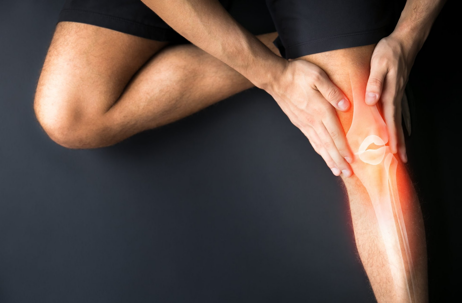
Anatomy of the Knee


The knee is an important joint comprised of three bones:
the Tibia, femur, and patella. The Tibia is also known as
the shin; the femur is the largest of the three bones and
helps to form the thigh; and the patella is known as the
kneecap. There is a fourth bone, the fibula which sits
next to the tibia and can sometimes play a role in knee
problems.
Cartilage covers the ends of the bones where they come
in contact with each other. This is called articular
cartilage. A second type of cartilage rests between the
femur and tibia. This is called the meniscus and it acts as
a shock absorber.
Ligaments are connective tissue that hold two bones
together. The main ligaments in the knee are the
cruciate ligaments that are in the center of the joint and
form a crisscross. They are the anterior and posterior
cruciate ligaments. There are also ligaments on each
side of the knee. One on the outside of the joint is the
lateral collateral ligament and the one on the inside of
the knee is the medial collateral ligament.
The joint space is contained within material called the
synovial. There is fluid within the joint called synovial
fluid which allows a smooth movement of the knee.
Muscles allow movement of the knee. The main muscle
groups in the knee are the quadriceps muscle which allow
the knee to straighten and the hamstrings which allows the
knee to bend.
Other components of the knee include tendons and joint bursa.
There are several knee conditions that can occur when one or
more of the anatomical components is not functioning properly.
If there is damage to the cartilage of the knee, arthritis can occur
and cause pain. Ligamental injuries are common in people who
play sports. Meniscus tears ca occur with wear and tear or with
injury.
It is important to seek medical care and advice with persistent
knee pain.


Leave a comment